BY RAEGAN NEUFELD
PHOTOS BY NICK McCOY
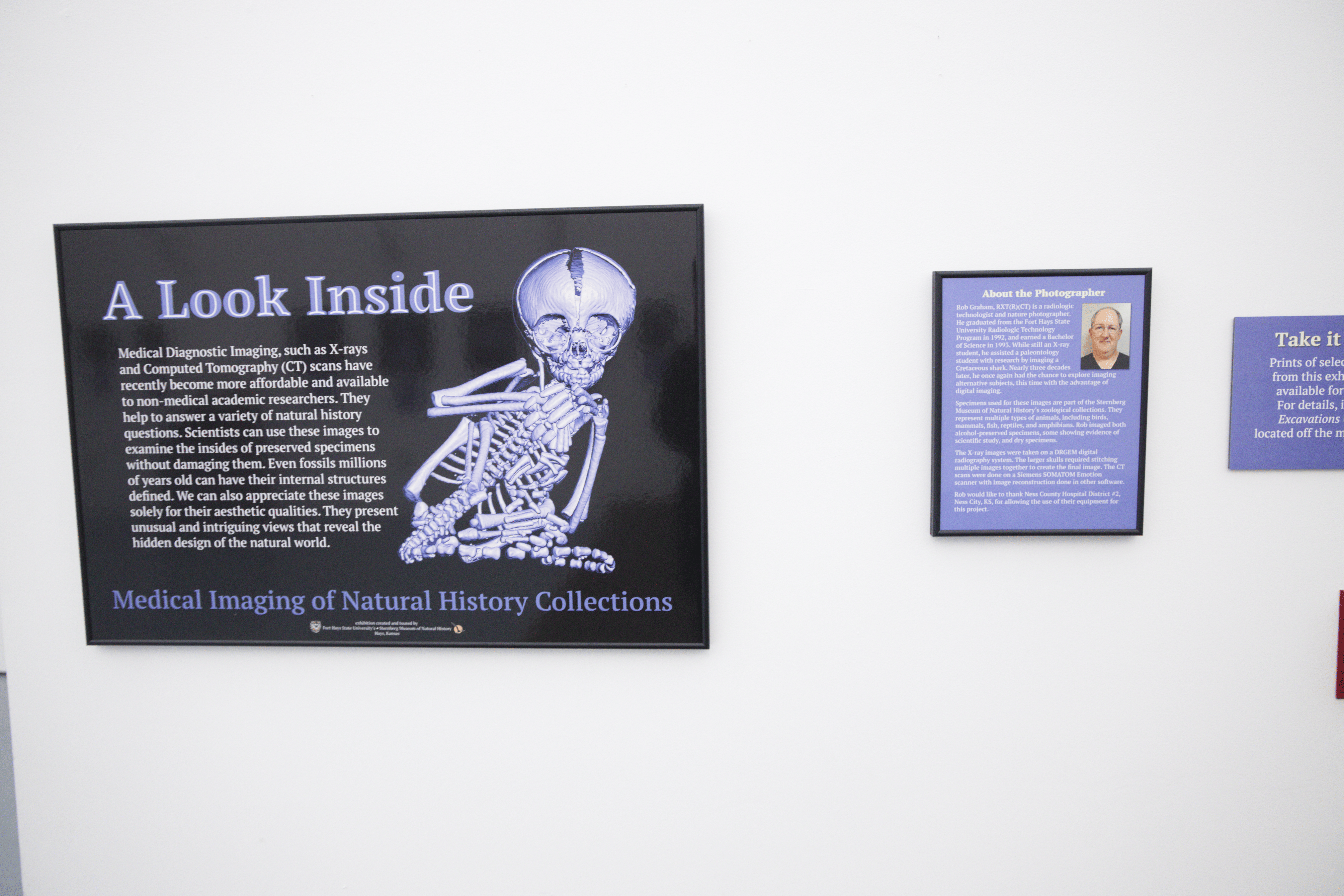
The Sternberg Museum of Natural History utilized its zoological collections and area medical technology to produce “A Look Inside,” an exhibit currently on display featuring X-ray images of various specimens.
According to museum director Reese Barrick, the exhibit was the brainchild of the late Curtis Schmidt who was the zoological collections manager from 2011 to 2022. Schmidt worked together with Rob Graham, a radiologic technologist, and Gregory Walters, the museum’s exhibits director, to capture the X-ray images and what information would go along with each one.
“This is kind of like [Schmidt’s] legacy now,” Barrick said. “A cool thing that he basically created.”
Some images are of whole specimens, such as reptiles and amphibians, stored in ethanol-filled jars, while others show the skeletons of similar animals or the skulls of birds and mammals. Some of the jarred specimens, skeletons and skulls are on display so patrons can compare them with the images.
“It’s a really fun way to get to see the secrets of things you can find out in nature, out in the Great Plains,” Barrick said.
Barrick also mentioned patrons rarely get to see a majority of the specimens on display.
“A lot of these things you don’t get to see because they’re on exhibit all the time. . . this allows you to take a look inside our collections and get an idea of the different cool things we can learn about,” he said.
A majority of the specimens come from Kansas, but some come from Nebraska, Oklahoma and Colorado. A few are even exotic, coming from other countries and had previously been at a zoo. For example, one X-ray shows the skull of a South American tapir.
“A Look Inside” is one of two current exhibits on display produced by the museum that will eventually travel to other places in the state. The other is “Grandeur in this View,” which shows microscopic images of bones and teeth. For “A Look Inside,” Barrick said there is more work to be done before it can be shipped anywhere.
“We’re working on trying to create good packing and display cases so we can send the actual specimens,” he explained.
Both exhibits will be on display until May 20. The museum is also working on another traveling exhibit, which will show the state symbols of Kansas, like the sunflower, tiger salamander, box turtle and more.
“They all have a natural history story,” Barrick said.
More information about current and upcoming exhibits, as well as hours and admissions information, can be found on the museum’s website.


You must be logged in to post a comment.